Duodenum
The duodenum curves around the pancreatic head.
It is located at L1-L3 level.
The vertebral level serves as an important landmark in locating the doudenum on a CT/MRI scan.

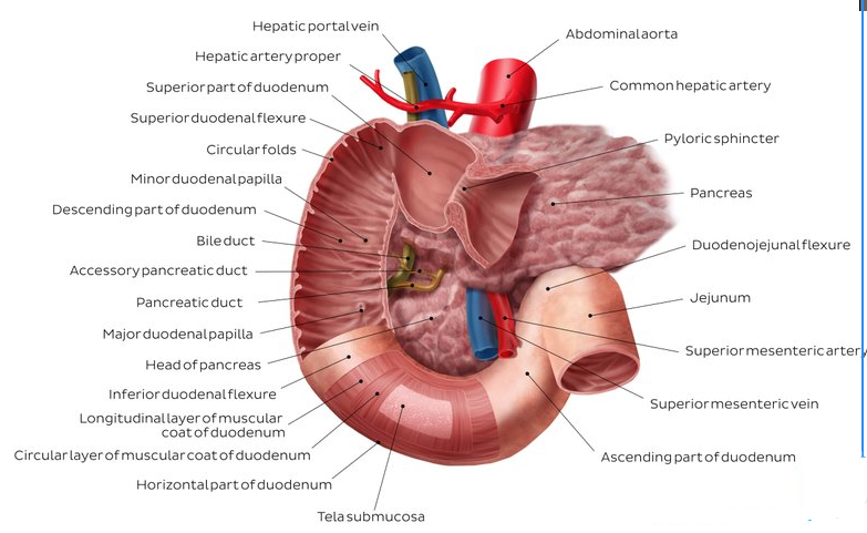
It is divided into four parts:
- Superior part. The stomach is an intraperitoneal organ as is the first part of the duodenum. The first part of the duodenum contains the duodenal cap, a dilation of the proximal duodenum that is easily identifiable on radiographs.
- The descending part and the rest of the duodenum lie retroperitoneally. The descending part courses deep to the transverse colon and anterior to the right kidney. The common bile duct enters its posterior wall.
- Within the duodenal wall, the common bile duct receives the main pancreatic duct (of Wirsung).
- Immediately after the junction, there is an enlargement called the major duodenal papilla (ampulla of Vater). The papilla is surrounded by smooth muscle called the sphincter of Oddi. An accessory pancreatic duct (of Santorini) may enter the duodenum proximal to the main pancreatic duct.
- At the opening there is an elevation of the mucosa, the major duodenal papilla (=papilla of Vater). Many people have an accessory pancreatic duct which empties into an additional papilla, the minor duodenal papilla (=papilla of Santorini).
- The horizontal part turns left and courses horizontally across the inferior vena cava, the aorta, and the vertebral column. In addition, the superior mesenteric artery and vein course anteriorly to the third part of the duodenum.
- The ascending part runs cranially along the left side of the vertebral column anterior to the aorta at the L2 vertebral level.
- This last part of the duodenum joins the intraperitoneally lying jejunum at the duodenojejunal flexure. Here the duodenum is attached to the back of the abdominal wall through the suspensory ligament of duodenum (=ligament of Treitz). Clinically the ligament of Treitz marks the border between the upper and lower gastrointestinal tract.
 .
.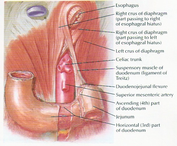
Ligament of Treitz, actually not a ligament at all, but the suspensory muscle of the duodenum, which participates in GI motility
- The distal portion of the fourth part is transitional, from retroperitoneal to intraperitoneal in the region of the duodenojejunal junction
The duodenum receives its blood supply from branches of both the celiac trunk (superior pancreaticoduodenal arteries) and the superior mesenteric trunk (inferior pancreaticoduodenal arteries).
| Innervation | Celiac plexus, vagus nerve |
Question
A 65-year-old man is admitted to hospital with vomiting. The vomitus is bilious.. A CT scan reveals that a large vessel is compressing the third (transverse) portion of the duodenum. Which of the following vessels is most likely involved in the obstruction?
The correct answer is E.
13—E: The superior mesenteric artery (and vein) course over the third portion of the duodenum.